Roberta Buiani and Gary Genosko
Abstract
In 2011, Australian-based artist Maria Fernanda Cardoso exhibited her multi-part work It is not Size that Matters, it is Shape at ARC One gallery in Melbourne. It was the first of a series of installations focusing on the reproductive organs of the Harvestman, a spider native of Southern Australia. Working in the tradition of natural history, Cardoso hoped to add new specimens to her collection, turning it into a “Museum of Reproductive Morphology.”
However, It is not Size that Matters, it is Shape exceeds the function of the natural history museum. Both the research methods that led to the exhibition and the choice of display of the organs reflect a novel ecological approach: the work is presented as a collaborative operation involving taxonomists, microscopists, graphic designers, and 3D printer professionals; the focus of the display is on the reproductive (or intromittent) organs of a single species using different formats, materials and scale. Upon facing the artifacts that compose Cardoso’s installation, one realizes that the hierarchies and taxonomies that traditionally define the natural world gradually blur and dissolve, as our anthropocentric approach to the non-human (both natural and fabricated) is both revealed and surpassed.
This text consists of two complementary parts: a critical commentary and an interview with Maria Fernanda Cardoso. These two writing forms (an academic paper and a dialogue) pay tribute to the multiplicity of voices that her installation evokes. In addition, they address both the artistic and scientific objectives of her work. The critical essay reflects on the diverse, intertwined issues explored by the artist, while the interview juxtaposes the voice of the artist with the interviewer’s interpretation, revealing how cultural threads and scientific tropes are woven together and intersect within scientific and artistic contexts.
Reassembling the Puzzle
In our visually-dominated culture, articles in scholarly journals and magazines in independent and mainstream media are accompanied by images and other visual reminders illustrating and completing their diverse textual content. Science is not immune from this tendency. The rise of scientific visualization and 3D modeling to both illustrate scientific outcomes to a scientific audience, and to popularize them for the lay public has encouraged lab technicians and graphic designers to develop innovative visualization techniques, nourishing a continuous quest for increasingly advanced and aesthetically original approaches to the processes of visualization.1
The variety of scientific illustrations that grace the covers of popular science magazines such as Scientific American or New Scientist and scholarly journals such as Cell or Nature are as much appealing as incomplete. In most cases the illustrations are the products of specific research and, as such, they seek to call attention to well-defined aspects that characterize the object of study, rather than to an entire phenomenon. In some cases, they are produced with the assistance of chemical or digital processes: when research items are submitted to the electron microscope, chemical preparation is needed to protect and better expose them to strength of the electron beam; the results are digitized and further processed using a number of ad hoc software. The resulting illustration will only emphasize those aspects that the experiment, by virtue of its own nature and use of materials, was meant to bring forth. We, the lay observers, are almost never aware of the unavoidable incompleteness hiding behind the mesmerizing images staring at us from the covers of journals (and thus of the different objectives that these images serve); nor can we identify the series of processes needed to produce these images. Would becoming aware of the processes used to obtain these images help us better understand the objects portrayed? Would displaying the entire set of processes in the same space – say, in a gallery — disclose the object analyzed in its “completeness” and reveal it in its biological and primordial purity (a sort of Kantian “thing-in-itself”)? Or would it rather change the naturalistic assumptions dominating the visual display of such object (that is, our assumptions that what we see is how the object looks exactly in nature)? Could this move initiate, for instance, a shift in the way we think of the entities measured, classified and portrayed by means of technological instruments and software? Should we, then, rather rethink of these objects as dynamic and hybrid substances, or, as artist Tagny Duff suggests, as inevitably entangled with the organic and the inorganic, the latter a legacy of the technologies and the modes of representation used?2
A multi-part installation conceived as a collaborative project at the intersection between science and the visual arts, Maria Fernanda Cardoso’s It is not Size that Matters, it is Shape asks similar questions. Cardoso’s installation is like a reassembled puzzle: it combines different techniques (2Dprints) and objects (3D printed sculptures) that abstract, digitize and re-materialize in different fashions the intromittent organs (the genitalia) of the Harvestman, thus revealing the multifaceted and diverse nature of this seemingly simple subject. In turn, with their ability to zoom into specific, yet always-incomplete aspects of their object of inquiry, this rich ensemble of techniques and sculptural and visual renditions reveal the limits, as well as the potentials of the scientific instruments of visibility. Finally, because of the particular topic Cardoso focuses on, this work is not limited to examining the multifaceted nature of scientific objects, but exposes how the absence, the refusal of science to confront the topic of sex, demonstrates its intrinsic and intimate relation to popular culture.
Cardoso locates her work in the tradition of nineteenth-century naturalists, characterized by the popular proliferation of (Victorian) scientists and amateurs interested in understanding nature through observation and collection of specimens (anatomical parts or botanical details, and the reproduction of natural artifacts through drawings and other visual techniques. Thus, her work can be defined as a “science and art project,” with science coming first, because of the research accuracy that this work implies, the extensive collaborative component, and the scientific material she uses.3 In fact, to produce these artifacts, the artist had to become familiar with the techniques routinely used by entomologists and microscopists to better observe and study specific morphological details proper of Harvestman spiders. In order to operate the instruments needed to produce these visual and sculptural objects, she had to partially rely on the expertise of these scientists.
With this installation, Cardoso fits and exceeds the scientific realm and the artistic/naturalistic tradition she is channeling. While single renditions draw attention to distinct aspects of the intermittent organ, none of them provides a comprehensive understanding. By exhibiting electron-micrographic scans, 3D models, and sculptural reproductions of the same object of inquiry (the intromittent organs of the Harvestman spider) side by side in the same gallery space, Cardoso shows how each of these techniques, in its own specialization, is incomplete. Each technique is only able to produce a partial portrayal of the object under scrutiny. Yet, when contemplated together in the gallery space, the artifacts resulting from these techniques instigate reflections on the specific knowledge and on the potentials that each of them claims, or is able to provide. Thus, her contribution is not only scientifically accurate, but it also takes on a critical role.
Thus, in laying bare the composite nature of scientific representation and visualization processes, this installation – along with an expanded body of work that incorporated other invertebrate specimen of both sexes recently exhibited at the Sidney Biennale and renamed as the Museum of Copulatory Organs”4 – does more than reveal the complementary nature of the artifacts on display. Her collaboration with scientists required that she mixed laboratory and gallery aesthetics, and that she dressed the objects into a gallery-appropriate form (i.e. she used top notch material for printing, and respected the display conventions of the exhibition space), yet without distorting or covering the scientific significance with over-aestheticized details. This is a delicate operation of balancing the two cultures. The objects on display expose the extent to which the minuscule reproductive organs of the Harvestman are manipulated and digitalized. The different states, seen together, induce questions on what is human-made and what is, supposedly, natural; what portion of the organs portrayed can (or can still, or can ever) be considered organic; and what is non-organic.
The multiple encounters occurring throughout this installation reveal with great clarity how and to what extent culture equally infiltrates the liberal arts and science. The sexual stereotypes evoked by the particular subject on display unify the two disciplinary contexts by virtue of their identical clichés. Thus, if one can draw one preliminary conclusion from this multifaceted project, it is that it helps us illuminate some hidden convergences and complementarities of liberal arts and science, human and non-human, what is assumed as natural and what instead appears as constructed.
Translating across dimensions
It is not Size that Matters, it is Shape is an exercise in translation across several dimensions: technical, trans-disciplinary, and cultural. This penchant for translation can be regarded as one of the first-noticeable revelatory layers in Maria Fernanda Cardoso’s installation (Cardoso, 2011). Each piece consists of the same object (the genitalia of the Harvestman), which Cardoso meticulously reproduced every time differently, thanks to a number of distinct techniques and by means of disparate media. While each artifact reproduces the Harvestman genitalia, it does so in different fashions and from different perspectives, adding more details and potentially improving our understanding of the object. Dissected and abstracted from the bodies to which they belong, the tiny organs of these insects underwent a series of operations before being immortalized as images, as 3D models and as objects. First, they were subjected to the scrutiny of a Scanning Electron Microscope (SEM). This resulted in 2D images (electron-microscopic scans). Using information retrieved from this first operation, the organs were turned into large-scale 3D models; using a 3D printer, the data was then translated into three-dimensional, solid sculptures. The entire collection of magnified and differently-processed organs was relocated side by side in the gallery. As electron-microscopic scans installed in tin boxes, as 3D models displayed as posters, and as sculptural objects. These objects rest in glass receptacles whose fit equally reminds the viewer of test tubes or of specially designed glass condoms. [Fig. 1]

Figure 1. Maria Fernanda Cardoso, Intromittent Organs of Series of Tasmanian Harvestman, Opiliones, 2008-2009; resin, glass, metal. 28 x 6 x 6 cm approx. Edition of 5 + 2 AP
In order to produce this multi-part work, Cardoso studied the anatomy of insects, consulted research in taxonomy and worked closely with scientists and microscopists. The genitalia of the insects were translated into different states thanks to a set of technologies that treat the specimens by protecting them with thin layers of special material (usually volatile metal) or by preparing them through the application of specific chemical recipes. This operation of protection helps the specimens endure the various processes of imaging, magnification and digitalization. The installation turns laboratory processes into artistic practice by revealing contact points between the two and by transposing cultural concepts and assumptions into scientific findings and vice-versa through various strategies of display.
As the reproductive organ of the insect is separated from its body—a body packed with organs and other complex intricacies—it is flattened into a bi-dimensional impression (a microscopic scan). In a later incarnation, the same organ is turned back into a three-dimensional print (the 3D model) and into a sculpture (the 3D printer artifact). In this latter incarnation, the genitalia are reproduced as a solid object showing the superficial structure of the original. This resulting artifact is not just a magnified version of the original, but a morphologically analogous, yet semantically different, sculpture.
Reproducing multiple versions of the same object would appear to be a simple exercise in aesthetics. However, not only do these products generate and underline different aspects of the organ based on the technical capabilities of the specific medium used or the parameters set by the software of choice. Each of them also constitutes an always different, partial reproduction of the surface of the object. As versions of the same object, they can be interpreted as complementary pieces that together provide a comprehensive representation of the external morphology of the Harvestman genitalia, highlighting their intricacy and their patterns, and illustrating how each of the items is not just a self-contained artwork but is part of a complex tableau.
Size, form, and glass condoms
The packaging of scientific and technological processes into an artistic project locates It is not Size that Matters, it is Shape at the crossroads between printing and photography, microscopy, biology and visualization, disciplines whose intertwining and communicative threads can be detected by means of Cardoso’s intentional act of cultural and linguistic translation. The presence of popular and scientific tropes and cross-references detected (albeit understood differently) indicate a cross-contamination that even a distracted viewer cannot dismiss. The observer can choose to interpret the work from a number of different perspectives and identify the threads that make such cross-contamination possible.
Issues regarding the study of technoscience, popular perception, and cultural tropes are juxtaposed with one another, each of them independently contributing to the meaningful title and content of this exhibition. The topic, the content, and the title given to this particular installation resonate at both the cultural and scientific level. For instance, the title It is not Size that Matters, it is Shape might simply acknowledge the scientific practice of Australian Museum microscopist Sue Lindsay. Lindsay introduced Cardoso to the findings of taxonomist Glenn Hunt (whose works Cardoso often credits in her research) regarding impressive variations in the Harvestman male genitalia that apparently make this recently-classified species unique.5 However, our prominent popular fixations with sex and the dimensions of sexual organs tend to overshadow the research done by the late Australian taxonomist, prioritizing other more provocative non-specialist connotations. The title can be read as a tongue-in-cheek reference to a tendency in the popular imagination to focus on stereotypes regarding the size of sexual organs in general, and to antagonistically oppose size to shape in popular culture. The artist herself laments the uncomfortable reactions she receives when the topic of her research is revealed. In addition to being notoriously used to compare sexual attributes, the prominence of “size” and “shape” in the title of the installation reminds us of the uneasy relation between the two terms. These two parameters in fact constitute a requirement in the description and measurement of any object. The former can be associated with mass production and the latter with design. To the puzzled requests for clarification regarding her chosen topic, she replies that her project has more to do with science than with art and popular assumptions, as it draws inspiration from the findings of, “…[T]axonomists [who] have long known that genitalia is diverse, and they use it as a useful tool for species differentiation.”6 With her work, Cardoso tries to go beyond the superficial obsession with sex in Western culture, to focus on the “great diversity and complexity in genitalic structures across the animal kingdom,” and to explore “…the relationship between the morphology of animal genitalia, its evolution and evolutionary forces at play, and the aesthetics of its representations.”7 However, transcending the above sexual assumptions might not be possible, as scientific material becomes inextricably entangled in her installation.
When it comes to sexual anatomy, size and shape are classically understood in antagonistic terms, the first being associated with quantity, the second with “mere” quality. While the title of Cardoso’s work prioritizes shape over size, both scientifically (as per Hunt’s finding) and sexually,8 the latter appears nonetheless to be a complementary and equally important component in the scientific process. It is only by artificially magnifying the Harvestman’s genitalia that we are able to admire their form as well as their artistic display; in order to show these organs as sculptures, the sexual organs had to be magnified. [Fig. 2]
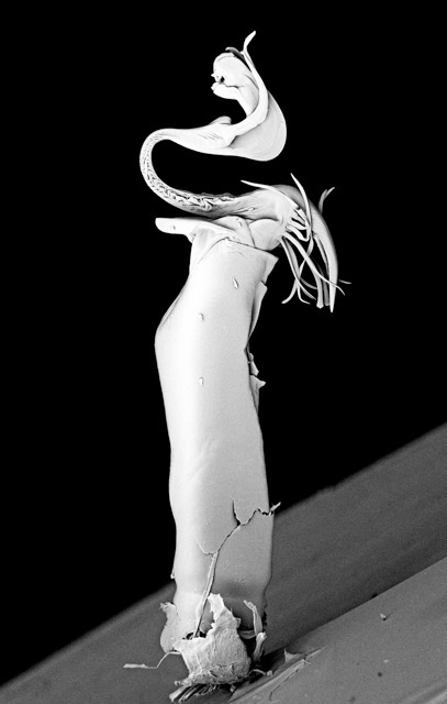
Figure 2. Electromicroscopic scan of the Thelbunus Mirabilius sp penis (Harvestman) Opiliones 2010. 31 x 24 x 4 cm mounted on tin box. Archival pigment print on 300gr cotton rag. Edition of 10 + 2 AP. From the collection of the Australian Museum SEM scan in collaboration with microscopist Sue Linsday.
Size and shape tap into the popular tendency to anthropomorphize non-human organisms. In this case, popular tropes of sexual power and male superiority could be easily ascribed to the Harvestman’s organs, thus establishing a direct link between the nature of invertebrates and that of mammals and human beings. As such, male domination (or a desire for it) seems to emerge from the gradual magnification of the genitalia by means of the micrograph and then through the 3D model and the sculptures. The latter evoke Darwinian reflections on sexual selection as a sub-category of natural selection: the differences in morphology of the Harvestman genitalia are either secondary sexual characteristics that have been studied by evolutionary biologists as evidence of post-copulation selection or the sign of a competition among males manifested through penis differentiation.9 In either instance, the female Harvestman spider may choose the male based on the morphology of its genitalia. The male penis determines its ability to attract the female, thus becoming a metonym for sexual talent.10
Beyond this interesting evolutionary finding, the representation of the genitalia in a sculptural and magnified form in combination with the anthropomorphizing reference to size and shape call attention to, and equally challenge, sexual and sexist stereotypes surrounding popular assumptions that domination obtains in size and hardness. In fact, the disembodied penises on display may be a magnified, marble-looking version of the original microscopic sexual organ. However, what makes them unique and intriguing is their morphology, not their size. Though their erect position evokes images of disembodied penises, the sculpted genitalia are inserted into clear glass receptacles that resemble condoms. While these repositories are there to protect the specimen from external damage, they can also be read as preventing them from spreading their ‘virtual’ semen. These penises are innocuous. Despite their brazen appearances, on a second look they project ideas of fragility rather than mighty power, as the glass receptacles are there either to keep them safe or to neutralize their potential.
The issue of size versus shape has a tendency to evoke anthropomorphic references and parallels between the life of insects (the Harvestman) and human beings, proper of a historical tradition that responds to a desire to understand in order to subjugate them. However, here the magnificent and very diverse growths springing from the magnified penises look more arborescent than human. This element speaks to a general interest in Cardoso’s body of work: her attraction to insects is not dictated by a drive to uncover the more obvious relation between the animal/insect kingdom and the human realm, as it transpires from many mainstream documentaries and commentaries – blockbusters such as Microcosmos11 and March of the Penguins12 come to mind. Her approach is more radical and to a certain extent anti-anthropocentric, as she points to evolutionary connections that all animals and insects share. Cardoso states in an interview, “There are about five philia (life form patterns) around this planet and somehow we all follow some of those basic architectures. Also I am particularly interested in mimicry strategies between plants and animals, I find it completely puzzling, and I think the art of copying is not exclusive to human culture but belongs widely in nature.”13 If seen from this perspective, the voluptuous mane topping the Harvestman penises reminds us that resemblances and interconnections should not be searched for in exclusively the human or the animal, but between all species.
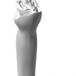
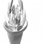
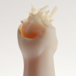
All the above elements may gradually shift the focus of the observer from popular tropes and sexual clichés to an increased attention to the aesthetics of the stunning diversity of these organs. Thus, the sculptures carry a double message, one that illustrates and provides evidence of scientific claims, and one that lays bare and dismantles popular myths of manhood and sexual power. [Fig. 3-5]
-
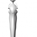
- Fig. 3 Intromittent organ of the Thelbunus mirabilus 2(Harvestman) Opiliones, 2011 Archival pigment print on 300gr cotton rag 196 x 87 cm, Edition 1/5 + 2 AP (front)
-
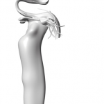
- Fig. 4 Intromittent organ of the Thelbunus mirabilus 1(Harvestman) Opiliones 2011 Archival pigment print on 300gr cotton rag 196 x 87 cm Edition 1/5 + 2 AP (side)
-
- Fig. 5 Intromittent organ of the Thelbunus Mirabilus (Tasmanian Harvestman) Opiliones, 2008-2009 resin, glass, metal. 28 x 6 x 6 cm aprox. Edition of 5 + 2 AP
Distributed natures
Across the exhibition space in wide variety, and of different scale and media, the Harvestman’s genitalia can be read as distributed objects in the sense that while there is no such thing as one single way to represent the Harvestman genitalia, the more objects are added to the installation, the more comprehensive a knowledge of these organs we will gather; while each object stands for a distinct aspect of the organ, it doesn’t reproduce it in its entirety; while each object can be read as an incomplete figure of the organ, yet it represents it;. In observing the alabaster-looking sculptures, the viewer is reminded of the sexual tropes evoked by those phallic objects even before wondering about their nature and origin, and finally about their location in the glass receptacles. Without reading the captions unveiling the identity of the object, the spectator has no context on which to draw. The aseptic, immaculate environment of the gallery and the conical glass test tubes containing the organs suggest that we are observing some oblong laboratory specimen with a varied morphology; their erect positions explicitly reminding the viewer of phalli. However, the observer’s ability to identify with certainty the Harvestman’s genitalia as “genitalia” is hindered by the separation of the organs from their original contexts, as well as by their disembodied state. The sleek superficial appearances and polished forms of these objects make us wonder whether they come from the natural and organic world or are rather the product of synthetic abstractions (for instance, from a bundle of data extracted from the digitalization realized by the process of microscopy and the microscope itself) arbitrarily transduced into material entities. What are they? Are they fossilized plants or other mysterious organisms? What is their real size?
The hypermediated nature of Cardoso’s objects makes us reflect on how conventions of display and technical procedures employed may affect the natural manifestation of the harvestman genitalia. The genitalia have been severed forever from the body of the Harvestmen so they can be properly examined; they have been frozen into a sculpture whose density and color mimic marble or alabaster, two dense, smooth, and rather solid minerals.
In most scientific contexts, images of microscopic organisms appear in journals and popular science magazines as isolated objects, devoid of the distraction that their surrounding context can pose to the viewer. Captions help us make sense of them only partially and we have no problem accepting the habit of displaying these scientific artifacts as isolated, disembodied objects; a “petri dish” approach that facilitates the work of the researcher, but that also inevitably de-contextualizes and neutralizes the object of study.14 The object appears to be turned into inert matter with connections to the external world that don’t exist or exist as pure mechanics.15 This approach leaves behind any element that would distract the viewer from sustaining a focused eye. Although it might be useful for utilitarian purposes, this purposely-partial approach confirms what Jane Bennett observes as an “image of dead or thoroughly instrumentalized matter” that “feeds human hubris and our earth-destroying fantasies of conquest and consumption.”16 Crucially, it prevents us from “detecting (seeing, hearing, smelling, lasting, feeling) a fuller range of the nonhuman powers circulating around and within human bodies.”17 Karen Barad echoes that this type of orientation ends up eliding (and thus trivializing and even ignoring) the “intrusion” of those social variables, such as sex, gender and race that allegedly belong to the exclusive sphere of the personal or individual.18
Cardoso’s display appears to generally abide by this scientific approach: she locates her objects in a quasi-laboratory, stereotypically aseptic setting. However, the genitalia of insects are anything but neutral objects. They cause reactions in the viewer that immediately call attention to the realm of culture. Upon a closer look, one realizes that this quasi-laboratory setting is strategically made to speak back to the viewer. For instance, Cardoso has decided to position her specimens vertically. This arrangement unavoidably calls back all those sexual topics that scientific display tends to ignore or avoids almost religiously. In this way Cardoso reclaims the right to make explicit those properties unfit or deemed marginal to a scientific context.
Cardoso claims to “explore different technologies and techniques according to the characteristics of the specimens.”19 In fact, the color of the erected sculptures resembles the neutral color (alabaster, or vero-white) obtained through the microscope (black and white). This strategy exposes the dominant techniques of visual rendition and modeling of scientific objects, and how the properties of these objects can be concealed, exposed or re-imagined differently according to the very techniques of display used. In other words, detached from the body of the Harvestmen, the genitalia become no different than any other object processed in the lab. Thus Cardoso’s work raises important questions regarding the scientific treatment of research objects.
On the one hand, the objects displayed in Cardoso’s work, sealed off and protected in their test tubes/condoms, look impeccably styled and polished as if sealed and isolated from any external impurity that might contaminate them. On the other hand, they look uncannily artificial (that is, of non-organic, non-natural derivation). The black-and-white rendition of the electron-microscopic scans, the monochromatic colors of the 3D model, and the semi-translucent vero-white color of the sculptures are equally the products of the materials and the instruments used, and the result of processes of magnification and clarification that the genitalia have gradually endured.
The scans were obtained using a Scanning Electron Microscope (SEM). Unlike light microscopes, the SEM shoots electrons to probe the surface of the specimen, generating black and white images as a result of the work of photons bouncing off the specimen. The resulting image is an exceptionally magnified three-dimensional impression of the object projected onto a two-dimensional support.20 The 3D model is a rendition obtained thanks to computer modeling software that processes the scan and extrudes the shape of the object according to specific parameters given by the designer. The 3D printer uses this data to rematerialize the object, or, as Cardoso states, to “make the object exist in the world again.”21 In fact, unlike sculpture and other manufacturing methods (like CNC, or Computer Numeric Control Machining), whose objects are obtained from removing material (subtractive process), 3D printing is an additive process comprised of laying down successive layers of material.
Cardoso might as well have assigned arbitrary colors to these artifacts, as it is customary in scientific visualization, but she opted not to. In fact, while the choice of color, or lack thereof, may remind the observer of the sanitized space of the laboratory popularized through TV series and films, its consistency locks the two-dimensional and three-dimensional objects together by unveiling their collective origin from a long-forgotten, dismembered insect. The latter aspects remind the viewer of the complementary qualities of the objects displayed in the gallery. Each of them, in fact, highlights always-different specific aspects of the genitalia, according to the medium and the process used. Cardoso credits a number of scientists and technicians as necessary contributors to this project. It is as if different artists – a painter, a sculptor, and a designer – collaborated in portraying the object using their medium of choice and their personal perspective. It is as if different scientists – a biologist, a pathologist, and a taxonomist – analyzed the same object, but read it differently every time according to the disciplines with which they are affiliated.
To obtain the objects in Cardoso’s work, the genitalia undergo a series of transformations over different volumetric stages: a three-dimensional biological organ becomes a three-dimensional shadow of the object projected onto a two-dimensional support. From there it is manipulated by software, its surface re-shaped as a three-dimensional solid object. During this transformation, the genitalia go from organic, to abstract, to inorganic; from biological/fluid, to digital, to solid/non-biological. In this alternative ecology, traditional classifications become obsolete. In fact, in the small universe of analogous (yet different) objects that Cardoso has located in the gallery, organic and inorganic, information and biology inherently coexist by folding into each other22 as each object composing the installation reproduces different aspects of the Harvestman’s genitalia.
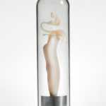
One might argue that the 3D printed object sitting in the glass test tube is only an imperfect copy of the superficial shape of the Harvestman’s genitalia. Yet, this object is an accurate rendition that in fact helps reveal and magnify the intricacy of the genitalia, rather than just describe it. The artifacts displayed in the gallery were obtained thanks to instruments that process the original digitally, turning its components into data. It is only then that the minuscule details of the genitalia can be extracted, magnified, and analyzed in their details. In order to restore the genitalia to a solid state, these data must be uploaded to software that re-combines and re-processes them first as a model, which is then extruded as a physical object to be printed layer upon layer. Thus, it would be partially incorrect to define the final object as an artificial version of the genitalia. It is, after all, composed by those very data from the original that are necessary to reproduce it in a much larger scale. In addition, the final product only exists thanks to the intervention of above processes. As Bennett among others argues, “a newfound attentiveness to matter and its powers… can inspire a greater sense of the extent to which all bodies are kin in the sense of inextricably enmeshed in a dense network of relations.”23 Thus, not only are the objects equally contributing to an understanding of the Harvestman’s genitalia. The objects are also inextricably joined together along with the viewers’ and the scientists’ respective assumptions, cultural baggage and biases. They form “a knotted world of vibrant matter, where to harm one section of the web may very well be to harm oneself.”24 Seen from this perspective, the object is neither a simulation nor a simulacrum, but a hybrid consisting of biological and digital information, an ecology in which discursive practices and the material world coexist. [Fig. 6-9]
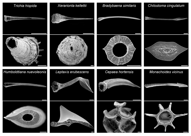
Figure 6. Base images to extrapolate forms for the models
-
- Fig. 7 Intromittent organ of the Allonuncia Grandis 1(Harvestman) Opiliones 2009 Archival pigment print on 300gr cotton rag 195 x 87 cm Edition 2/5 + 2AP Edition 1/5 + 2AP Trienal de Santo Domingo
-
- Fig. 8 Intromittent organ of the Allonuncia Grandis 2(Harvestman) Opiliones 2009. Archival pigment print on 300gr cotton rag 195 x 87 cm Edition 1/5 + 2 AP
-
- Fig. 9 Intromittent organ of the Allonuncia grandis Harvestman 2008-2009, resin, glass, metal. 28 x 6 x 6 cm aprox. Edition of 5 + 2 AP (Detail)
Science and Culture
Cardoso’s new ecology manifests beyond the hybrid and concerted materiality of the objects on display. It exposes how science and culture are always and necessarily woven together, yet are also arbitrarily kept separated. Using visual language, microscopy and the representation of scientific phenomena (via visualization and modeling) are fully embedded in a system that mixes and exploits technical information and “entertainment” on the one hand, and on the other hand well-established visual tropes and innovation aspiring to move away from these tropes. These aspects are often in opposition to each other. Yet they coexist any time the microscopic is reproduced. Cardoso mixes aesthetics and science, popular assumptions and information in her installation, revealing the artificiality of these sectorial divisions even when they are included in specialized or exclusive contexts, such as science fairs and art exhibitions, demonstrations geared towards a science audience, and popular science illustrations. Although Cardoso’s work can be read differently by unlike observers, it also creates a platform where science and culture intersect, enabling different observers to share analogous principles and references, imagining them as spectators and learners simultaneously.
Traditionally, the practice of observing organisms through a lens was an experience that combined a sadistic attitude towards dirty and base, yet marvelous and fantastic, organisms under scrutiny with the “exacting observation” of the rigorous scientific material of the invisible unknown where “…mysterious animalcules with unexpected powers could be not only watched under the enlarging lens, but controlled and directed, even as they rushed by.”25 Thus, the approach of the scientist towards the object seen through the microscope was more the one of a voyeur than a detached viewer. The installation successfully breaks an old, yet still surviving division “between a sensuous, pleasurable, or merely curious watching and a rational, tasking, language-driven observation [that] arose during the eighteenth century.”26 Rather than the entertaining nature of a scientific image portraying a microscopic organism (the “enjoyable watching”) distracting the viewer from the specific information (“exacting observation”) provided by the image, the entertaining component of “enjoyable watching” draws attention to the image, informed by a historical practice of seeing the object as the “spectacle of nature” and as the “work of god.” In selecting intromittent organs—a “scientific and sanitized term” for genitalia, a term already packed with allusions to sexual activity—as the objects of her installation, and in displaying them as artifacts in an exhibition, Cardoso lays bare and mixes the voyeurism implicit in watching and extracting scientific information (thus achieving scientific pleasure, a scientific form of porn) with the aesthetic—and a bit perverse—pleasure stemming from observing the exquisite organs on display.
Conclusion
As we explore the numerous links and cross-references between science, popular culture, and technological artifacts, we gradually become aware of the multifaceted nature of Cardoso’s artwork. In fact, the work invites us to reflect on the modalites and the roles of representation in science and the arts. Despite its scientific accuracy, It is not Size that matters, it is Shape is neither an example of visualization or scientific illustration, nor an exhibition of science education. Tagny Duff, a bioartist who has produced multi-part installations that address similar concerns as Cardoso’s, observes that the main goal of these composite projects “is not to produce or prove a theory or hypothesis, let alone to create a canon of artifacts and documentation.”27 Cardoso’s installation uses established techniques and scientific processes to produce multiple objects; she reproduces the obtained objects in an exceedingly exquisite fashion and displays them in a clinical, yet fashionable manner, according to the exhibition rhetoric appropriate to the venue. Thus, she doesn’t produce any explicitly new information about the objects on display.
As in the case for a number of other artists who have become involved in arts and science collaborations, Cardoso participates in and does not reject the conventional rules existing in the sciences and in the cultural rituals of looking. Being well-aware of the seductive power of the tropes that dominate her objects of research, she appropriates and exploits them, amalgamating them in an installation that equally combines scientific content and aesthetics. By doing so, she breaks these very rules and rituals. In fact, by displacing the objects from the lab and relocating them into the gallery, and by engaging the audience in debates about specific issues raised by their research, Cardoso encourages new reflection on the complexities and problematic elements perpetuated by cultural/scientific notions that these objects convey.
Thus, gathering and displaying such a body of work into one place reveals the impossibility of grasping the full meaning and importance of the scientific object by means of a single image or product; it shows how diverse techniques can interpret an object rather differently. In addition, the work illustrates how producing such a comprehensive installation may raise awareness regarding the amount of information and subsequent research that can be extracted from such tiny specimens. Thus, Cardoso’s work requires and appears to obtain an all-encompassing, holistic approach that acknowledges the deeply connected and multidisciplinary universe it unleashes, a rich ecology comprised of media, nature and culture complementing each other.28
Because of her use of biological specimens in a gallery environment, Cardoso’s body of work could be –erroneously – be compared to a plethora of other artists known as bioartists. Like Oron Catts and Ionat Zurr, Tagny Duff and Natalie Jeremijenko, to name a few, Cardoso creates, with her installation, a complex ecology consisting of an economy of practices, objects, and materials. Every piece of the multipart installation is a facet of a complex phenomenon, yet, it forms a whole when it is coupled with the other pieces on display.
Facing the variety of practices comprising the sphere of bioart, Robert Mitchell explains that the term “ought to encompass all those works of art that engage biotechnology in some way.”29 While Cardoso’s work employs biotechnological practices and instruments, her interest and goals lie somewhere else: she is in fact neither interested in intervening in the critical debate regarding the future of biotechnology (a prophylactic approach30), nor is she using biotechnology as a medium (a vitalistic tactic).31 Instead, Cardoso inserts her work in the tradition of the natural science museum, reproducing its classic display, attempting an augmented display of one specific object. In this exhibition, the audience may re-discover points of contact between popular culture and big science, and become aware of the fragmented way through which science communicates. Her work is not projected towards manipulation and speculation; it is simply about display and ecological representation. Finally, unlike most bioartists, whose final display in the gallery merely constitutes one phase (or the documentation, as Duff and Hauser put it)32 of a continuous biological process, Cardoso’s work focuses on the very objects on display. These objects are the final product, one that makes sense as a single artifact as well as as a whole, comprehensive and ensemble.
Cardoso’s latest installation testifies to her loyalty towards the natural museum display and her belief in the object. In the summer of 2012, Cardoso participated in the 18th Sydney Biennale (June 27-Sept. 16) with an exhibition inspired by It is not Size that matters, it is Shape entitled MoCo (the Museum of Copulatory Organs). This new work incorporated a much larger and more diverse collection of objects. It included samples of both male and female genitalia of invertebrates, reproduced through a wider range of technological techniques and modes of display. In addition to the already mentioned 3D modeling, 3D printing and electron-microscopy, Cardoso deemed glass the ideal medium to recreate naturally transparent organs and contacted a glass artist in order to reproduce spermatophores.
With the increased quantity and variety of objects on display, Cardoso appears to have broadened her focus. Her work seems no longer concerned with questioning perceived tensions between science and culture, art and science, size and shape, but is more broadly concerned with exploring and dispelling assumptions regarding sexual organs of insects and animals in general. To accommodate the richer collection, Cardoso ditched the aseptic white walls for settings reminiscent of a typical natural history museum. MoCo is installed in a dark space, where spotlights illuminate original natural science specimens, vero-white 3D prints, 3D models, glass sculptures, and scans collected in showcases. The ambiguity that dominated the location of the previous installation does not dissipate, but simply shifts from the location in which the viewer was temporarily left uncertain as to whether the exhibition space belonged to a laboratory or a gallery, to the displayed object itself. Insects, specimens, and other items (for instance, pollen) are re-sized and placed side by side simultaneously acting as specimens and as artworks. Despite Cardoso’s evolving interests, her research parabola and conceptual understanding of the material she manipulates are still tuned to the same approaches to ecology.
For a Museum of Reproductive Morphology, A Dialogue with Maria Fernanda Cardoso
Q. Can you tell me about your training in the use of microscopes, the use of which were vital for your new project on the intromittent organs of Harvestman ‘ticks’?
A. Hola…. I have no training, but my dad got a really good quality loupe scope for himself and two for my sister and I when we were kids; we took them to the bush to observe nature. I remember looking at fungus that would release spores when you pressed on it. For this project, however, I approached the Australian Museum Electromicroscopy Unit and have been working with microscopist Sue Lindsay. A Harvestman is a Daddy Longlegs with short legs; actually, it’s not a spider, and not a mite, but in-between, with a family of its own, the Opiliones.
Q. Why did you select the Harvestman for your studies of intromittent organs?
A. When I first started with Sue, rather than start by dissecting insects looking for the genitalia, I dealt with genitalia that had already been prepared. She had worked with mites and Harvestmen in collaborations with a taxonomist, the late Glenn Hunt.33 And she had all the files. She found the specimens and we started scanning. We also worked with the Phallomedusa solida, the genitalia of a snail recently named after the shape of its phallus, and the specimens were in the collection. Harvestman penises are relatively easy to remove and prepare, which is why taxonomists use the genitalia to help identify different species. The insects look very much alike, but not when you examine their private parts, which are quite varied. Hunt also wrote a paper about some really long penises he found. I only worked on one, but didn’t use it in my series, since the image was not great. Further, even if Harvestman penises are easy to dissect, they are the size of a speck of dust and you can barely see them with your eyes. But when they are placed under the microscope, you see these elaborate shapes. It is mind-blowing.
Q. Where did the Harvestman samples come from?
A. Tasmania.
Q. Glenn Hunt’s scientific papers on the Harvestman credit Sue Lindsay and Roger Springthorpe for the SEM – Scanning Electron Microscope images – and line drawings, respectively. Did you learn how to use the scanning electron microscope? Or does Sue make the images under your direction?
A. No, that’s a specialized job. I sit next to Sue and direct. At home I also have to look at hundreds of images we take and choose from them, then have them photo-shopped by a professional so the quality of the image is just right. I sit next to him, Nick Greenwich, who is also my printer, and we work together on the desktop and I tell him what I want. By the way, the print quality is top notch, on beautiful papers, utilizing a 12-ink DaVinci printer, on very thick cotton rag paper. They are ink prints and the paper absorbs the ink … exquisite. At University of Sydney microscopy lab they tried to train me to use their new MicroCTScanner, but I have no patience to spend a million hours in front of a computer and it’s not my language; so, they have been helping me, too, although in theory students get trained and rent access to their equipment.
Q. The “hypertrophied male genitalia” paper by Glenn and an Argentine colleague raises some interesting possibilities – does size really matter? I suppose the scientific answer is that it depends entirely on adaptation – sexual selection – of the female genitalia that receives it – very long vaginas.
A. There are a bunch of evolutionary biologists studying genitalia and developing the most interesting theories. Some consider the shape and intricacy a form of post-copulation sexual selection, where the female chooses (cryptic female choice) not only prior to mating but afterwards. Some others consider genital morphology a form of sexual war, and in other cases a form of sexual coevolution; sperm wars are also brought into play as some genital appendages are designed to remove sperm from previous mating, the shape is about competing with other males as females are promiscuous in general (in most species with some exceptions).
Q. Are you going to consider female genitalia?
A. Yes, I have imaged some “epigynum” of female spiders but need more specimens so I can have more diversity. They consist of two spirals. Male spider palps are also spirals. He actually breaks the tip of his pedipalps inside her in order to block her from releasing his sperm.
Q. Scientific representation is an important and understudied element in scientific discourse and communication. How did the SEMS and line drawings influence or inform your own decisions in remaking the penises in different media?
A. I wanted to make 3D objects as I am trained as a sculptor and I have problems with so many 2D images in the world that already exist. I believe objects have a presence and capacity to engage us differently than 2D images. For the Harvestman I explored computer modeling and 3D printing, and it was incredibly hard but really rewarding. Making objects is about making them exist in the world again, at a different scale, but sharing our space. But some specimens are not suitable for objects and the image alone is important. I consider that I need all modes of representation depending on the specimen, so I like diversity of life, and diversity of representation. I am also using other techniques, such as bronze, glass and drawings, video, light microscopy, etc. so my project is diverse, like nature is, and it depends on what suits each specimen best.
For example, I have made models of damselfly genitalia (looking at illustrations from WG Eberhard’s Sexual Selection and Animal Genitalia). I used a new technology called bronze clay, which you work as if it were clay, and then fire it in a kiln in a bed of coals; it turns out to be 99.9 pure bronze. The reason I like it is because it shrinks about 15-20% so the detail is superior to working in wax and casting it in bronze.
Q. I’m curious about your choice of container for the finished sculptures – not exactly a test tube, but a lab-like container in a rough bullet shape. Is the base of the sculpture metallic? How did you decide on this mode of presentation? They don’t look like laboratory glassware!
A. I had them made by a renowned glass artist in Canberra, Peter Minson – the long skinny ones on top of the objects. I wanted the glasswork to look like condoms to emphasize the shape of the genitalia! We first tried using glass glowing, but it was too hard to make them all the same. As a result it’s done with glass tube and flame. Apparently it’s really hard to make them. Only a very experienced glassmaker can do a job of that quality. The bases were also made to fit the glass. They are just protection from dust and contact, but I actually can show them without the glass so you can see the detail up close. I have also worked with flame glass artist Mark Elliot from Sydney to make pseudoscorpion spermatophores. He and I shared an interest in the exquisite XIX C glass models of Leopold Blaschka and his son, Rudolph (especially the Harvard glass flowers and marine invertebrates).
Q. The microsculptures are ethereal. The ‘skin’ colour is provocative. The shapes display – at least in photographs – movement quite well, even in the container. Can you explain why you chose the hint of human skin tone?
A. It’s 3D printing, so my choices were a semi-translucent or that vero white.I don’t see them as skin colour but more like alabaster white, as they are translucent a little bit like a marble. To me, they are reminiscent of the white on black SEM images. I didn’t want paint or electro plate or anything like that on the models as the detail is so fine that you don’t want anything on top diminishing the detail. I think the “skin” colour is in the viewers’ minds, not in the objects.
Q. Have you embedded these studies of genitalia into the art history of genital representations? Human or otherwise?
A. I will! But I will avoid humans; there is too much pornography in the world. I am interested in things people have never seen, and that are “weird and wonderful.” I am making a museum of Reproductive Morphology and this body of artworks are some of the specimens in the collection! I am hoping to launch it at the Sydney Biennale in June 2012 and we are in the process of looking for funding. Depending on the results, it – the collection – will be bigger or smaller. My passion is for invertebrates, although some marsupials are worth looking at, and a few mammals. But those will be last. I am working in the tradition of natural history collecting and taxonomy, so I can keep adding up specimens one by one, growing the museum collection slowly but surely.
- Dolores Steinman and Steinman, David, “Toward New Conventions for Visualizing Blood Flow in the Era of Fascination with Visibility and Imagery,” ed. Oliver Grau and Thomas Veigl (Cambridge MA: MIT Press, 2011), 129–148 ↩
- Tagny Duff, “Going Viral: Live Performance and Documentation in the Science Laboratory,” Performance Research 14, no. 4 (2009): 36–44 ↩
- STUDIOchannel132, “18th Biennale of Sydney – Maria Fernanda Cardoso,” July 2012 ↩
- Artscape, “Professor Maria Fernanda Cardoso and the Museum of Copulatory Organs,” April 2011 ↩
- G. S Hunt, “A New Cavernicolous Harvestman From Western Australia (Archnida: Opiliones: Triaenonychidae),” Australian Journal of Entomology 11, no. 3 (September 1972): 232–236 ↩
- 18th Biennale of Sydney – Maria Fernanda Cardoso, 2012. http://www.youtube.com/watch?v=6J1srEtZr-I&feature=youtube_gdata_player ↩
- STUDIOchannel132, “18th Biennale of Sydney – Maria Fernanda Cardoso” ↩
- G. S. Hunt, “Hickmanoxyomma, a New Genus of Cavernicolous Harvestmen from Tasmania (Opiliones: Triaenonychidae),” Records of the Australian Museum 42, no. 1 (March 1990): 45–68 ↩
- Charles Darwin, The Origin of Species (Gramercy, 1995) ↩
- Hunt, “Hickmanoxyomma, a New Genus of Cavernicolous Harve” ↩
- Nuridsany, Claude, and Marie Perennou. MicroCosmos, 1996. http://topdocumentaryfilms.com/microcosmos/ ↩
- Jacquet, Luc. March of the Penguins Documentary, 2005 ↩
- “Maria Fernanda Cardoso: Small Wonders,” Australian Art Collector, June 2011 ↩
- Roberta Buiani, “Visualizing as Exorcism: Learning from Viruses” (Singapore, 2008), 134–138 ↩
- Jane Bennett, Vibrant Matter: A Political Ecology of Things (Duke University Press Books, 2010) ↩
- Bennett, Vibrant Matter, ix ↩
- Ibid, vii ↩
- Karen Barad, Meeting the Universe Halfway: Quantum Physics and the Entanglement of Matter and Meaning (Duke University Press, 2007) ↩
- “Maria Fernanda Cardoso: Small Wonders” ↩
- John J. Bozzola and Lonnie Dee Russell, Electron Microscopy : Principles and Techniques for Biologists (Sudbury, Mass.: Jones and Bartlett, 1999); Edward K. Wagner and Martinez J. Hewlett, Basic Virology (Malden, MA: Blackwell Science, 2004) ↩
- See Interview below ↩
- Anna Munster, Materializing New Media : Embodiment in Information Aesthetics (Dartmouth, NH: Dartmouth College Press, 2006) ↩
- Bennett, Vibrant Matter, 39 ↩
- Ibid, 40 ↩
- Barbara Maria Stafford, Good Looking : Essays on the Virtue of Images (Cambridge, Mass.: The MIT Press, 1996) ↩
- Barbara Maria Stafford, “Voyeur or Observer?: Enlightenment Thoughts on the Dilemmas of Display,” Configurations 1, no. 1 (1993): 95–128 ↩
- Duff, Tagny. “Going Viral: Live Performance and Documentation in the Science Laboratory.” Performance Research 14, no. 4 (2009): 36–44 ↩
- Félix Guattari, Chaosmosis : an Ethico-aesthetic Paradigm (Bloomington, Ind.: Indiana University Press, 1995) ↩
- Mitchell, Robert, Bioart And The Vitality Of Media (In Vivo) by Robert Mitchell (Seattle: University of Washington Press, 2010): 22 ↩
- Ibid., 27 ↩
- Ibid., 28 ↩
- For the processual aspects of bioart as performance and documentation and the failure of the exhibition space to contain them see Duff, Going Viral, and Jens Hauser, Bio Art – Taxonomy of an Etymological Monster. (UCLA Art/Sci Center series, 2006) ↩
- The papers of Glenn S. Hunt that I consulted were “Revision of the Harvestman genus Miobunus from Tasmania,” Records of the Western Australian Museum Supp. 52 (1995): 243-52; idem and Emilio A. Maury, “Hypertrophy of male genitalia in South American and Australian Triaenonychidae,” Memoirs of the Queensland Museum 33/2 (1993): 551-56; Hunt Revue Suisse de Zoologie ↩